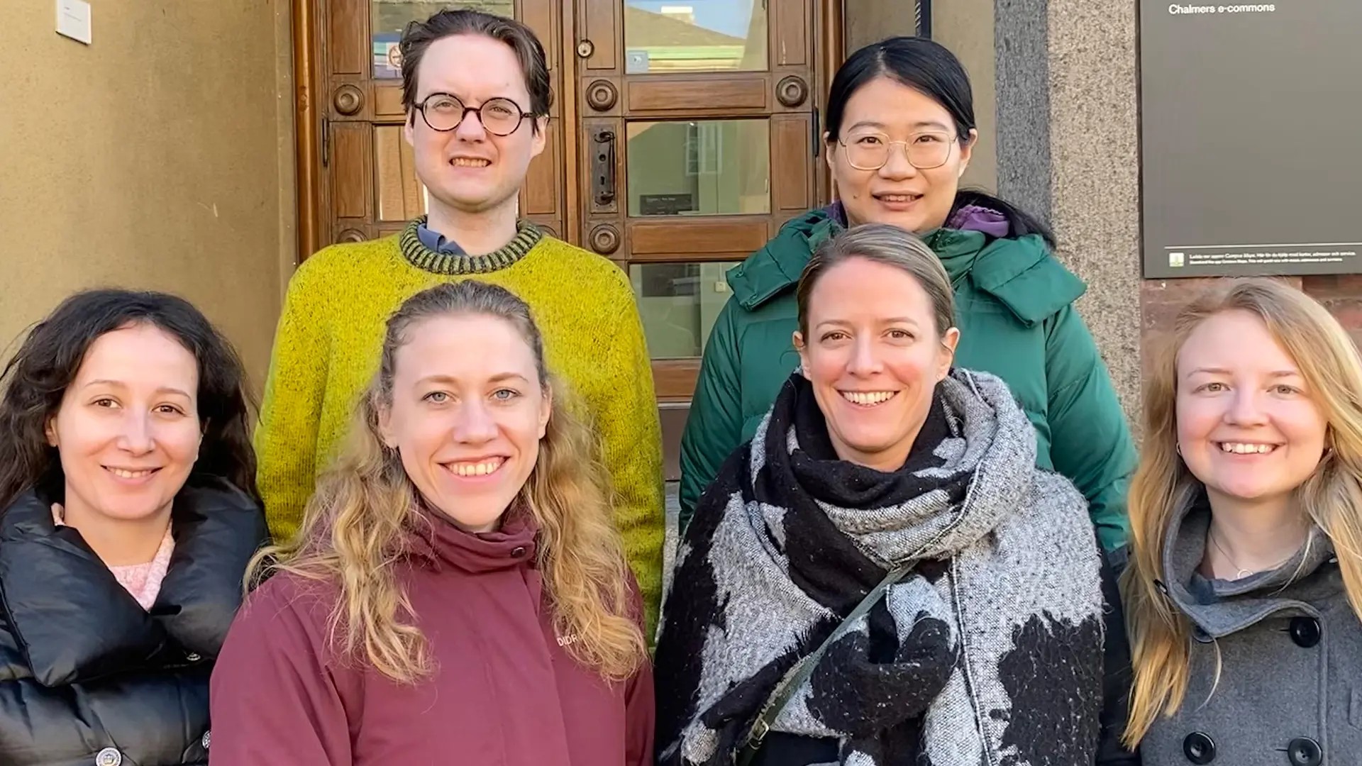
The focus of our research is in the development of X-ray scattering and imaging techniques and their application towards materials with hierarchical structure.
About the group
The applications we are working on in different collaborations are spanning a broad range from biomimetic hierarchical nanocomposites, materials based on cellulose, industrial injection-molded plastics as well as the characterization of bone and soft tissue. A common denominator of these diverse applications is the arrangement of nanometer-sized building blocks within macroscopic samples, in particular the alignment of anisotropic constituents. To that end, SAXS tensor tomography, which allows to look at nanostructure alignment in 3D macroscopic samples is a powerful tool. We perform our experiments at different synchrotron beamlines in Europe, and we collaborate closely with the Swiss Light Source at the Paul Scherrer Insitute (Switzerland) where part of the research group is located (SMAM Group, PSI).
News
Narwhal tusk research featured in video game
Exciting to see that some of our research on Narwhal tusk made it into an educational videogame about climate change in the Arctic and its impact on some of its inhabitants!
The game “The Tipping Point” was developed by Fabula studios and can be downloaded for free at https://fabulastudios.dk/the-tipping-point/
If you make it over collapsing ice sheets to level 10 and 11, you will get to the part which is based on our interdisciplinary research project with Chalmers University of Technology, Aarhus University, and the Greenlandic Institute of Natural resources, funded by Nordforsk.
You can use the “Tusk Analyzer 6000” on X-ray Fluorescence data to look at elemental mapping within growth layers of the tusk.
Release of Mumott 1.0
We are happy to announce the release of MUMOTT 1.0, an all-Python package for the analysis of tensor tomography measurements! The package employs a modular framework with several choices for representation, optimization and regularization. It also includes support for GPU-accelerated computations. The package is available via PyPI and can be installed with python -m pip install mumott. For more information, see the documentation at mumott.org. Doi link from Zenodo: https://zenodo.org/record/8070314
Research projects
Unravelling the spiral structure of narwhal tusk
Narwhals are mysterious animals whose tusk has a unique spiral structure with highly anisotropic mechanical properties. In this project, we use 2D and 3D SAXS imaging, birefringence microscopy, X-ray fluorescence and advanced electron microscopy to study the orientation and degree of anisotropy of the biomineral nanoparticles in the tusk in 2D and 3D. Examining samples from dentine and cementum layers will reveal the unique structure and shed light on its formation process. The study of the 3D hierarchical structure is very valuable for micromechanical modelling of anisotropic composites, which will bring new knowledge on composite materials with complex architectures and inspire new material designs.
Dimitra Athanasiadou
Development of methods and theory for tensor tomographic reconstruction
The computation of tensor tomographic reconstructions is a problem that involves large datasets and a large number of computations. The development of improved algorithms and more optimized code is therefore an important aspect of this research. In addition, an improved understanding of the theory and mathematics behind tensor tomography is necessary for more efficient experiments, as well as for developing reconstruction methods further and generalizing methods from small-angle X-ray scattering to e.g. systems involving visible light. The project is funded by the Swedish research council (VR 2018- 041449) and the European research council starting grant MUMOTT (ERC-2020-STG 949301).
Leonard Nielsen
X-ray imaging for characterization of pharmaceutical formulations
Pharmaceutical formulations can be utilized as a method for controlled release, for improving dissolution properties and to enhance the bioavailability of an active substance. A key issue to understand and design the functional mechanism of these formulations is to understand its structure and morphology. In this project we explore how one can apply different x-ray imaging and scattering techniques to investigate the structure from micro to nanoscale and to relate the functional properties of the compound to its morphology.
Martina Olsson
Structuring of fibrillar systems under flow studied with SAXS
Controlled alignment of anisotropic materials, for example cellulose nanofibrils is of high interest, since their mechanical properties are highly dependent on the orientation. The fluidic four-roll mill (FFoRM) device provides an easy way to study any arbitrary 2D flow by small angle X-ray scattering (SAXS) or birefringence imaging. Knowledge gained on flow behaviour can then be used for process development. The project is related to the development of the ForMAX beamline at MAX IV, which will be a combined SAXS and imaging beamline, in particular aimed at the Swedish forest industry.
Marianne Liebi
Bio-based materials structure
Forest raw materials are promising sustainable materials for a variety of applications. This project focuses on the applications of forest raw materials in both soft-matter (achieving anisotropic in fluidic four-roll mill device) and energy storage (to be integrated into energy storage device). Advanced characterizations, e.g. birefringence microscopy, X-ray scattering and imaging techniques, will be implemented to investigate the microstructures of the bio-based materials to further understand the mechanisms. The work is part of Wallenberg Wood Science centre, the Treesearch collaboration platform and the MAXIV facility with the ForMAX beamline.
Andi Di
Using X-rays to characterize conventional polymers to wood-based thermoplastics
Investigation of the structure of polymer materials is of interest since it defines the mechanical properties of the material. For many applications, understanding the structure is key in order to prevent breakage, increase the quality of the material, and develop more sustainable materials. In this project we use X-ray based imaging to characterize synthetic and cellulose-based polymer materials to relate how processing, structure and mechanical properties are related.
Linnea Björn
SAXS Tensor Tomography Workshop
Periodicly, a SAXS Tensor Tomography Workshop is organised. The first edition was held at the Department of Physics on February 2020 with participants from Chalmers, Lund University, University of Surrey and NTNU Norway. This first eddition was focused on experimental set-up and practicalities during the experiment, real problem solving during data analisys and data visualisation.
Open positions
For new Student projects, Master thesis, PhD and Postdoc positions contact the group leader Marianne Liebi.
Previous projects
Publications
Group members
Research group leader
Post docs
PhD students
Contact
- Affiliate Docent, Materials Physics, Physics


