


Aktuellt


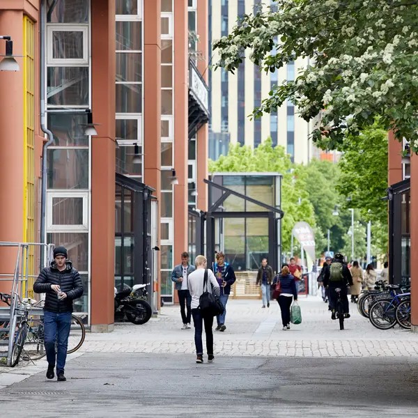
För en bättre framtid


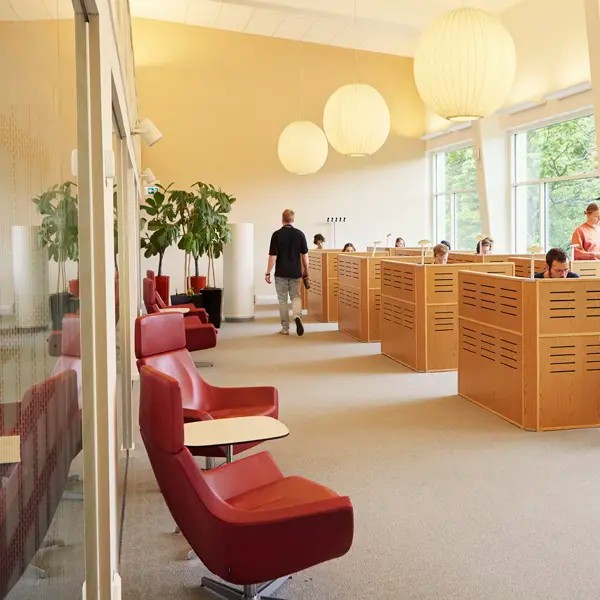
För dig som studerar på Chalmers
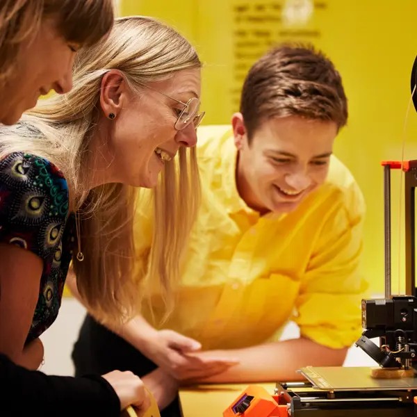


Tillsammans utvecklar vi framtiden


Chalmers i siffror (2023)
- 10 773helårsstudenter
- 3 026publikationer/år
- 977doktorander
- -25,8%minskade klimatutsläpp (2019-2023)

Varje dag driver vi viktiga samhällsfrågor framåt, testar nya idéer och bidrar till framtidens utveckling.